Adhesions are bands of scar-like, fibrous tissue that can form when there is any kind of tissue injury. According to Van Den Beukel et al. (2017), adhesions can cause pelvic pain. They also reports that “reformation of adhesions has been linked to relapse of pain after adhesiolysis” (Van Den Beukal et al., 2017). Hermann and Wilde (2016) note that adhesion formation is “highly prevalent in patients with a history of operations or inflammatory peritoneal processes”. (Endometriosis is an inflammatory disorder.)
Hao, M., Zhao, W. H., & Wang, Y. H. (2009). Correlation between pelvic adhesions and pain symptoms of endometriosis. Zhonghua fu chan ke za zhi, 44(5), 333. Retrieved from https://pubmed.ncbi.nlm.nih.gov/19573306/
“Conclusion:Pelvic adhesions are characteristic lesions of endometriosis, the site and degree pelvic adhesions are closely correlated with pain symptoms.”
Abd El-Kader, A. I., Gonied, A. S., Mohamed, M. L., & Mohamed, S. L. (2019). Impact of endometriosis-related adhesions on quality of life among infertile women. International Journal of Fertility & Sterility, 13(1), 72. Retrieved from https://europepmc.org/article/med/30644248
“The prevalence of adhesions resulted from endometriosis was 37.6%. Demographic characteristics of the women with endometriosis-related adhesions were not significantly different from those of women without endometriosis- related adhesions. The most common location for endometriotic adhesions was adnexal adhesion (51.2%) followed by adhesion of anterior abdominal wall (24.4%). Quality of life was significantly impacted by endometriosis related adhesions (P=0.002).”
Lee, Y., Lee, Y., Lee, S., Jung, S., & Chon, S. (2020). Correlation of preoperative biomarkers with severity of adhesion in endometriosis. Journal of Gynecology Obstetrics and Human Reproduction, 49(1), 101637. Retrieved from https://www.sciencedirect.com/science/article/pii/S2468784719301217
“Preoperative blood Serum and CA 125 results were obtained and pelvic adhesion scores were calculated. The patient group with adhesion scores less than 28 points was defined as the mild adhesion group, and those with a score of 28 or more were members of the severe adhesion group. The CA 125 level was significantly higher in the severe adhesion group than in the mild adhesion group. The CA 125 level, size of the largest cyst, and WBC count were associated with the level of pelvic adhesion. Adhesion scores were significantly higher in the CA 125 ≥ 35 U/mL group than in the CA 125 < 35 U/mL group. Patients with a preoperative CA 125 level higher than 35 U/mL are at high risk for pelvic adhesion.”
“The negative impact on the tubo-ovarian unit can be directly by distorting the anatomy, indirectly by invoking inflammation or by oxidative damage with poorer-quality oocytes. Endometriosis even seems to have a negative effect on pregnancy outcome after in vitro fertilization.”
Van Den Beukel, B. A., de Ree, R., van Leuven, S., Bakkum, E. A., Strik, C., van Goor, H., & ten Broek, R. P. (2017). Surgical treatment of adhesion-related chronic abdominal and pelvic pain after gynaecological and general surgery: a systematic review and meta-analysis. Human Reproduction Update, 23(3), 276-288. Retrieved from https://academic.oup.com/humupd/article/23/3/276/3058801
“Experts do not classify endometriosis as an autoimmune disease. However, endometriosis may increase a person’s risk of developing an autoimmune disease, as well as other chronic conditions. The reason for the link is unclear, but it might exist because endometriosis causes inflammation, which may contribute to an imbalanced immune response.
“An autoimmune disease is one in which the body mistakenly attacks its cells, tissues, or organs. The resulting damage can cause a wide variety of symptoms, depending on which part of the body it affects. The abnormal immune response that occurs in endometriosis may be due to an existing autoimmune disorder. The evidence is not clear as to which condition causes the other.
“There is still no conclusive cause of endometriosis, and researchers do not yet know what triggers the condition. However, abnormal immune system responses and genetics may be among the factors that play a role in the development of the disorder. A person with endometriosis may also have an increased risk of comorbidities. Comorbidities are conditions that exist alongside a primary condition….
“Treatment for an autoimmune disease typically focuses on suppressing the immune system so that it stops attacking healthy cells in the body. Endometriosis does not appear to respond to any known treatments for autoimmunity.”
“While the most commonly seen symptoms of the disease are pelvic pain, dysmenorrhea, and infertility, endometriosis has also systemic effects in multiple organ systems. Here, we review literature describing closely associated comorbidities including cardiovascular disease, cancers, autoimmune disease, psychiatric conditions, and metabolism/body weight. We examine the pathophysiology and hypothesized mechanism by which endometriosis may lead to these systemic effects; mechanisms include cytokine and micro-RNA production as well as stem cell migration and dissemination. The broad systemic effects of endometriosis as well as correlated comorbidities are often overlooked in the treatment of patients with endometriosis. Increased awareness may lead to more effective treatment and prevention.”
Endometriosis close to the urinary organs, like the bladder, can cause symptoms such as pain with urinating (dysuria), blood in the urine (hematuria), urinary frequency/urgency/incontinence. However, it is important to note they may NOT cause symptoms. This is important because endometriosis around the ureters (the tubes that take your urine from your kidneys to your bladder) may not cause symptoms but can lead to kidney failure. Close follow up with your provider is important.
Bladder:
Symptoms are similar to interstitial cystitis (see “Interstitial cystitis“) and may include (potentially cyclical but not necessarily so) urgency, suprapubic pain, pain with urination, blood in the urine, inflammation of the bladder lining, etc.
Ferrero, S., Bogliolo, S., Menada, M. V., Ragni, N., Biscaldi, E., Camerini, G., & Remorgida, V. (2009). Diagnosis and management of bladder endometriosis. Journal of Endometriosis, 1(3-4), 113-121. Retrieved from https://doi.org/10.1177/2284026509001003-401
“Bladder endometriosis is defined as full-thickness infiltration of the detrusor; small sub-peritoneal implants and small nodules of the vesicouterine fold cannot be considered to be bladder endometriosis. In women with endometriosis, urinary tract involvement is rare (1% to 5% of cases) but the bladder is affected in 80% to 84% of these cases. Symptoms of bladder endometriosis are various and not specific: besides pain symptoms, patients may complain of urinary frequency, urgency, urge incontinence, dysuria, and hematuria. Although bladder endometriosis may be suspected at vaginal examination, the preoperative diagnosis is based on transvaginal ultrasonography and magnetic resonance imaging. Medical therapies may temporarily reduce the severity of symptoms related to the presence of vesical endometriosis; however, the symptoms may persist in cases of large bladder nodules or may recur after cessation of therapy. Surgery represents the gold standard for treatment of bladder endometriosis and laparoscopy should be preferred to laparotomy. Excision of bladder nodules may be performed either by partial-thickness resection or by partial cystectomy according to the size and depth of the infiltration of the lesions in the bladder wall. Persistent improvement of symptoms has been demonstrated at long-term follow-up, particularly when the lesions involve the vesical dome.”
Akpınar, S., Yılmaz, G., & Çelebioğlu, E. (2015). A rare cyclic recurrent hematuria case; bladder endometriosis. Quantitative imaging in medicine and surgery, 5(3), 485. Retrieved from http://www.amepc.org/qims/article/viewFile/4389/5294
“…urinary tract involvement especially the bladder endometriosis is a rare entity in women of reproductive age with clinical symptoms of cyclical urgency, hematuria and suprapubic pain. We herein present magnetic resonance imaging (MRI) findings of spontaneous bladder endometriosis case with cyclical hematuria symptoms.”
Ureter endometriosis, while rare, is important for a provider to assess. It does not have many specific symptoms to identify and so can be insidious with its harm. The studies below highlight endometriosis in other areas that have been shown to occur frequently with endometriosis of the ureters.
“Little attention has been paid by the renal literature to ureteral endometriosis, a rare and silent disorder that can eventually lead to renal failure. In endometriosis, the ureteral involvement can be limited to a single ureter, more often the left one, or both ureters with consequent urine tract obstruction and ureterohydronephrosis. In most cases, the ureteral obstruction is caused by endometrial tissue surrounding the ureter (extrinsic ureteral endometriosis). In the remaining cases, endometrial cells are located within the ureter (intrinsic ureteral endometriosis). Progressive ureteral obstruction can be insidious in onset and can ultimately lead to renal failure if a correct diagnosis is missed. The true incidence of renal failure caused by endometriosis is completely unknown, although cases have been reported in the literature. The diagnosis of ureteral endometriosis is difficult since the disease may be clinically silent or associated with non-specific symptoms. Only a high index of suspicion and radiological support may help to obtain an early diagnosis. However, while renal imaging is useful in the cases of extrinsic endometriosis, the diagnosis of intrinsic endometriosis often requires ureteroscopy or laparoscopy. The prognosis of ureteral endometriosis depends on the time of diagnosis. In too many cases of bilateral obstruction, the patient is referred to the nephrologist because of an advanced, irreversible renal failure. Although some patients may benefit from progestin or anti-arotamase therapy, in most cases of ureteral endometriosis surgery is needed, laparoscopy surgery being preferred today to laparatomy.”
Nezhat, C., Paka, C., Gomaa, M., & Schipper, E. (2012). Silent loss of kidney seconary to ureteral endometriosis. JSLS: Journal of the Society of Laparoendoscopic Surgeons, 16(3), 451. Retrieved from https://www.ncbi.nlm.nih.gov/pmc/articles/PMC3535807/
“Ureteral endometriosis is a serious localization of disease burden that can lead to urinary tract obstruction, with subsequent hydroureter, hydronephrosis, and potential kidney loss. Diagnosis is elusive and relies heavily on clinical suspicion as ureteral endometriosis can occur with both minimal and extensive disease. Surgical technique to treatment varies, but the goal is to salvage renal function and decrease disease burden.
“Although a relatively common gynecologic condition, localization to areas distinct from the peritoneum, ovary, and rectovaginal septum occurs in up to 12% of women with endometriosis.3 Pelvic endometriosis can infrequently involve the urinary tract system in approximately 1% of cases, which is a prevalence of 3.5 million women worldwide.4 The bladder is the most commonly involved site and the urethra the least. Of these localizations of disease, ureteral endometriosis accounts for approximately 10% of genitourinary involvement, which is 350,000 women worldwide.4,5 In endometriosis, ureteral involvement is often limited to one ureter, commonly the left, and can potentially lead to urinary tract obstruction, ureterohydronephrosis, and loss of renal function. There are estimates that 30% or nearly a 100,000 women with ureteral endometriosis will have 25% to 50% loss of nephrons at time of diagnosis of ureteral endometriosis, and an unknown number will then have loss of the kidney.6 This final insult of complete loss of renal function is exceedingly rare.
“Ureteral endometriosis is a serious localization of disease burden. Asymmetric involvement of endometriosis, with the left pelvis more commonly involved than the right, is readily explained by anatomic differences of the pelvis.12 The distal segment of the ureters and bladder are the more frequently involved locations due to the proximity of the reproductive organs.13 Additionally, ureteral endometriosis is more likely to be associated with rectosigmoid lesions as opposed to bladder involvement.14 Two major pathological types exist: extrinsic and intrinsic ureteral endometriosis. In the extrinsic type, which is the most common, endometrial glandular and stromal tissue involve only the adventitia of the ureter or surrounding connective tissues, whereas the intrinsic type involves the muscularis propria, lamina propria, or ureteral lumen.1”
Kidney/Renal:
Giambelluca, D., Albano, D., Giambelluca, E., Bruno, A., Panzuto, F., Agrusa, A., … & Lagalla, R. (2017). Renal endometriosis mimicking complicated cysts of kidney: report of two cases. Il Giornale di chirurgia, 38(5), 250. Retrieved from https://www.ncbi.nlm.nih.gov/pmc/articles/PMC5761639/
“Although usually occurring in pelvic organs, endometrial lesions may involve urinary tract. Renal endometriosis is extremely rare and it has only occasionally been reported in the past. We report two cases of patients with renal cystic lesions, incidentally found at imaging techniques during oncologic follow-up for gastric sarcoma and melanoma, initially misinterpreted as complicated haemorrhagic cysts and then histologically characterized as renal localizations of extragenital endometriosis.”
Endometriomas are a type of endometriosis cyst on the ovary. Management of endometriomas can be complex as there are many schools of thought on how they should be handled. Generalists, gynecologists, or fertility experts will often suggest a wait-and-watch approach when faced with a patient who has an endometrioma. Some will suggest that surgery is only warranted for cysts above a certain size. They might even cite potential damage in the context of fertility concerns. A watch-and-wait approach is a reasonable option for many and sometimes even a skilled excision surgeon will recommend the same, depending on your circumstances. The trouble is, unlike many other kinds of cysts (the common kinds that are not related to endometriosis), no amount of waiting will change the fact that endometriomas do not resolve on their own. There are different techniques used when surgically treating an endometrioma. Some will just drain the cyst, but that doesn’t eliminate what caused the cyst to form in the first place. Even with effective skilled excision, other endometriomas can present later on. Accessing more advanced surgical care makes sense for two key reasons: (1) removing an endometrioma effectively is a challenging task and (2) endometriomas can be an indicator of more extensive endometriosis elsewhere that will also need to be addressed.
“Ovarian endometriomas are indicators for pelvic endometriosis and are rarely isolated. Particularly, left endometriomas were found to be associated with rectal DIE and left uterosacral ligament localization and bilateral endometriomas correlated with adhesions and pouch of Douglas obliteration, whereas no correlation was found between endometrioma size and DIE. Determining appropriate management, whether clinical or surgical, is critical for ovarian endometriomas and concomitant adhesions, endometriosis, and adenomyosis in patients desiring future fertility.”
“Endometriomas (ovarian endometriotic cysts) are a commonly diagnosed form of endometriosis, owing to the relative ease and accuracy of ultrasound diagnosis. They frequently present a clinical dilemma as to whether and how to treat them when found during imaging or incidentally during surgery. Previously published guidelines have provided recommendations based on the best available evidence, but without technical details on the management of endometriosis….Owing to the limited evidence available, recommendations are mostly based on clinical expertise….It is generally accepted that endometriosis presents in three different entities, which are frequently found together: peritoneal lesions, deep endometriosis and ovarian endometriotic cysts (endometriomas) (Nisolle and Donnez, 1997). Endometriomas are probably the most commonly diagnosed form of endometriosis because of the relative ease and accuracy of ultrasound diagnosis. Although their exact prevalence and incidence are not known, they have been reported in 17–44% of women with endometriosis (Busacca and Vignali, 2003). The presence of ovarian endometriomas has been reported as being a marker for deep endometriosis (Redwine, 1999) and multifocal deep vaginal, intestinal and ureteric lesions (Chapron et al., 2009).”
“The objective of this study was to evaluate the significance of severe preoperative pain for patients presenting with ovarian endometrioma (OMA)…. After multiple logistic regression analysis, uterosacral ligaments involvement was related with a high severity of chronic pelvic pain [odds ratios (OR) = 2.1; 95% confidence interval (CI): 1.1–4.3] and deep dyspareunia (OR = 2.0; 95% CI: 1.1–3.5); vaginal involvement was related with a higher intensity of lower urinary symptoms (OR = 13.4; 95% CI: 3.2–55.8); intestinal involvement was related with an increased severity of dysmenorrhoea (OR = 5.2; 95% CI: 2.7–10.3) and gastro-intestinal symptoms (OR = 7.1; 95% CI: 3.3–15.3). CONCLUSIONS: In case of OMA, severe pelvic pain is significantly associated with deeply infiltrating lesions. In this situation, the practitioner should perform an appropriate preoperative imaging work-up in order to evaluate the existence of associated deep nodules and inform the patient in order to plan the surgical intervention strategy.”
“”Not all cystic lesions in the female pelvis are ovarian. It is important to consider disease processes that may mimic those of the ovaries because they affect patient management. It also is important to be familiar with the imaging characteristics of the various pelvic cysts, their anatomic locations, and the patient’s clinical history to make an accurate diagnosis……..peritoneal inclusion cyst, paraovarian cyst, mucocele of the appendix, obstructed fallopian tube (eg, hydrosalpinx, pyosalpinx, and hematosalpinx), uterine leiomyoma, adenomyosis, spinal meningeal cyst, unicornuate uterus, lymphocele, cystic degeneration of lymph nodes, lymphangioleiomyomatosis, hematoma, and abscess. A cystic pelvic mass is nonovarian if it is separate from the normal ovaries.”
The term thoracic endometriosis has been used to describe the varying clinical and radiological manifestations associated with the growth of endometrial glands and stroma in the lungs or the pleural surface. Catamenial pneumothorax (CP) is defined as pneumothorax happening around the menstrual period and is the most common manifestation of thoracic endometriosis, accounting for about 80% of cases. In almost all cases, thoracic endometriosis is unilateral and right sided, although there are rare cases of left sided disease. Bilateral disease is extremely rare. The presentation of CP includes cough, chest pain, and shortness of breath. The chest pain may be similar to patients with spontaneous pneumothorax or present as shoulder, scapular or neck pain.
Some management strategies for thoracic endometriosis are as follows. For any women with a spontaneous recurring pneumothorax, a gynecologic history and evaluation of her menstrual cycle should be done. When thoracic endometriosis is suspected, Video-Assisted Thoracoscopic Surgery (VATS) is the preferred approach. The diaphragm needs to be explored thoroughly, including the visceral and parietal pleura, and if necessary, a port should be inserted at the subcostal margin to assess the posterior diaphragm. All accessible lesions and fenestrations should then be resected. Fulguration or ablation of lesions should not be used, as it is inadequate for treatment of endometriosis and will result in higher recurrence rates. Following the resection, plication is recommended to seal and strengthen the diaphragm. Simple suturing of the fenestrations does not provide tissue diagnosis and is usually followed by recurrence. Lesions close to the phrenic nerve or its main divisions are best treated by limited resection (if possible) and repair. A mechanical pleurodesis is also further recommended after all accessible lesions have been excised.
We also propose a joint surgery by the thoracic surgeon and the gynecologist specialized in endometriosis wherever possible. Not only will this allow for an assessment of the diaphragm from both the pleural and peritoneal side concurrently, this will also allow more thorough identification of endometriotic lesion and fenestrations by both specialists. Additionally, there is a significant association between the presence of pelvic endometriosis and thoracic endometriosis and a joint surgery will further allow the treatment of any pelvic endometriosis at the same time.
Medical treatment has long been considered the first choice in patients with thoracic endometriosis. The literature contains a variety of reports on the use of oral contraceptives, progestational drugs, danazol and gonadotropin-releasing hormone (GnRH) agonists. Experience in the last three decades has been greatest with danazol and GnRH agonists. However, the results of medical treatment for CP have been disappointing. At 6 and 12 months, surgical treatment of CP resulted in far lower recurrence rate than did hormonal therapy (5% and 25% compared with 50% and 60%). Therefore, before initiating pharmacologic disruption of ovarian steroid genesis in a young woman, all surgical treatment options should have been exhausted.
While endometriosis of the thoracic area is rare, it can occur. Several case reports and studies that are cited below, report symptoms of coughing up blood (hemoptysis), isolated chest pain, and/or shortness of breath with menstrual cycles. At times, a collapsed lung (pneumothorax) has been reported (symptoms such as shortness of breath, chest pain, fast or shallow breathing). One study demonstrates that thoracic endometriosis has been reported since earlier than 1968! There are also links for further information as well as to a facebook group for extra pelvic endometriosis.
“Conclusion: The diagnosis and management of thoracic endometriosis requires a multidisciplinary approach, based upon skillful differential diagnosis, and involving careful gynecologic evaluation and assessment of the cyclicity of pulmonary symptoms. Imaging findings are non-specific, though there may be laterality towards the right lung. Since symptom recurrence is more common in those with presenting with pneumothorax, post-operative adjuvant medical therapy is recommended.
“…Thoracic endometriosis is defined as the presence of ectopic endometrial tissue inside the thoracic cavity [8]. It usually presents with pneumothorax, hemothorax, hemoptysis, lung nodules, isolated chest pain or pneumomediastinum; symptoms are synchronized with the menstrual cycle [5]. A pneumothorax occurring between 24 hours before and 72 hours after the onset of menstruation is described as “catamenial.” Although this may include primary spontaneous pneumothorax, occurring coincidently during the perimenstrual period, the majority of recurrent episodes of catamenial pneumothorax are caused by thoracic endometriosis. It is encountered in 20% to 30% of women with spontaneous pneumothorax [9].”
“Catamenial pneumothorax (CP) is generally caused by intraperitoneal air leaking from the uterus into the thoracic cavity via a defect in the endometrial tissue of the diaphragm and is usually detected in the right thorax. We report a case of left-sided CP caused by endometriosis in the visceral pleura and with no abnormal findings in the diaphragm. A 33-year-old female patient presented at the end of a course of low-dose contraceptive pills for pelvic endometriosis, with spontaneous pneumothorax in the left chest. Chest CT revealed a bulla in the left upper lung lobe.”
“Thoracic endometriosis syndrome is a rare disorder characterised by the presence of functioning endometrial tissue in pleura, lung parenchyma, airways, and/or encompasses mainly four clinical entities–catamenial pneumothorax, catamenial haemothorax, catamenial haemoptysis and lung nodules. The cases were studied retrospectively by reviewing the records at Amrita Institute of Medical Sciences, for duration of five years i.e., form March 2010-2014 and analysed for the clinical presentation and management of thoracic endometriosis syndrome. Catamenial breathlessness was the main symptom. Pneumothorax and pleural effusion were the findings on investigations. Histopathology report of endometriosis was present in three cases (50%). Conditions with excess oestrogen like endometriosis, fibroid, adenomyosis were diagnosed in these patients by pelvic scan. After the initial supportive treatment with hormones, pleurodesis, hysterectomy and lung decortication were the treatment modalities. Two cases that had multiple recurrences were diagnosed as disseminated TES. They underwent combined treatment of surgery and hormones.”
Hwang, S. M., Lee, C. W., Lee, B. S., & Park, J. H. (2015). Clinical features of thoracic endometriosis: A single center analysis. Obstetrics & gynecology science, 58(3), 223-231. Retrieved from http://www.ncbi.nlm.nih.gov/pmc/articles/PMC4444519/
“A total of 21 patients were retrieved, 15 of which were eligible for inclusion. Of these 15, 8 patients were diagnosed with thoracic endometriosis with hemoptysis as their chief complaint, and 7 patients presented with pneumothorax. The median age was 35 years (range, 23–48 years). All patients displayed some degree of catamenial symptoms, although patterns differed; some patients reported symptoms with every cycle of menstrual bleeding (n=7), whereas others showed only occasional episodes during menstruation (n=8). Patients had experienced between 1 and 7 catamenial episodes before presenting for medical advice. None of the 15 patients were smokers; 1 patient had a history of asthma, and 2 patients presenting with catamenial hemoptysis had histories of empirical tuberculosis medication-use prior to being diagnosed with thoracic endometriosis. Three patients had previously undergone video-assisted thoracoscopic surgery (VATS), undertaken at other centers, following which they had not been diagnosed with thoracic endometriosis. In 1 patient with a history of pneumothorax (P12), the episode occurred on the contralateral side to the presence of endometriotic lesions and the underlying etiology was considered equivocal.”
“A 25-yr-old Caucasian woman was referred by her GP with a 3 yr history of right shoulder pain. The shoulder pain frequently occurred at the start of menses and was responsive to ibuprofen. Clinical examination, shoulder radiographs and blood tests (CRP, rheumatoid factor) were all normal. The MRI of the shoulder and right hemi-diaphragm showed small areas of high signal on T1, T2 and STIR sequences at the lateral right hemi-diaphragm, consistent with areas of recent haemorrhage, suggestive of ectopic endometrial tissue ( Fig. 1 ). Positive identification and ablation of the endometrial tissue by laparoscopy was thought to be a low-yield procedure so the patient commenced the continuous progestogen—only pill which caused amenorrhoea and resolution of her symptoms.”
“A 54-year-old woman, born in Hungary but living in Australia during the past eight years, was found on mass miniature radiography to have a shadow in the right lung and was referred for further investigation. She gave a history of exertional dyspnoea for the past few years, during which she had gained weight….There was no history of cough, haemoptysis, or chest pain. She had had two uncomplicated pregnancies, 35 and 14 years previously, and her menopause had occurred in 1964 with no subsequent bleeding. She gave no history of any operation or serious illness. INVESTIGATION A radiograph of the chest (Fig. I) showed a thin-walled cystic lesion in the right midzone with a little irregularity of the lower wall of the cyst.”
Nancy’s Nook is devoted to the individual with endometriosis. Some have fertility questions and concerns. Out of respect for all our members, we’ve compiled this list of online resources that those who are trying to conceive, who have conceived, or who have had children may find helpful. Please note that we are not able to judge how accurate the information in these groups is; that’s beyond our scope. We’ve just tried to point you in a direction that might be helpful to you.
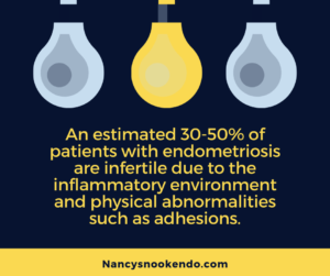
Infertility is strongly associated with endometriosis, and for some it may be the only symptom that they recognize (American Pregnancy Association, 2012). An estimated 30–50% of women with endometriosis have infertility (Macer & Taylor, 2012). Endometriosis can be “minimal” and does not have to be an advanced stage for it to affect fertility (Bloski & Pierson, 2008).
We have collected a few links and articles that address endometriosis and infertility that you can explore and better educate yourself to discuss with your providers. Infertility is a difficult journey that is highly individualized. There is no one correct answer.
Macer, M. L., & Taylor, H. S. (2012). Endometriosis and infertility: a review of the pathogenesis and treatment of endometriosis-associated infertility. Obstetrics and Gynecology Clinics, 39(4), 535-549. Retrieved from https://www.ncbi.nlm.nih.gov/pmc/articles/PMC3538128/pdf/nihms422379.pdf
“The association between endometriosis and infertility is well supported throughout the literature, but a definite cause-effect relationship is still controversial. The prevalence of endometriosis increases dramatically to as high as 25%–50% in women with infertility and 30–50% of women with endometriosis have infertility (2)…. Endometriosis affects gametes and embryos, the fallopian tubes and embryo transport, and the eutopic endometrium; these abnormalities likely all impact fertility. Current treatment options of endometriosis-associated infertility include surgery, superovulation with IUI, and IVF.”
“Endometriosis was associated with a greater risk of pregnancy loss (spontaneous abortion: RR 1.40, 95% CI 1.31–1.49; ectopic pregnancy: RR 1.46, 95% CI 1.19–1.80). Endometriosis was also associated with a greater risk of GDM (RR 1.35, 95% CI 1.11–1.63) and hypertensive disorders of pregnancy (RR 1.30, 95% CI 1.16–1.45).”
“Results showed that the prevalence of high uNK cells was 33.1%. Prednisolone significantly decreased the uNK cell concentration (P < 0.001), however reduction to normal limits was achieved in only 48.3% of patients. There was no difference in any of the pregnancy outcomes or complications between women who had received prednisolone and those who had not. In conclusion, this study showed a relatively high prevalence of raised uNK cells in women with recurrent reproductive failure and confirmed the effect of prednisolone on reducing uNK cell concentrations. We found however no evidence for a significant beneficial effect for prednisolone therapy on pregnancy outcomes. Until the results of an adequately powered RCT become available however, these findings should be considered preliminary.”
Nesbitt-Hawes, E. M., Campbell, N., Maley, P. E., Won, H., Hooshmand, D., Henry, A., … & Abbott, J. A. (2015). The surgical treatment of severe endometriosis positively affects the chance of natural or assisted pregnancy postoperatively. BioMed research international, 2015. Retrieved from https://www.ncbi.nlm.nih.gov/pmc/articles/PMC4515280/pdf/BMRI2015-438790.pdf
“Results. In the study period, 355 women underwent surgery for stage III-IV endometriosis. Follow-up data are available for 253/355 (71%) women. Postoperatively, 142/253 (56%) women attempted to conceive with a conception rate of 104/142 (73%). Confidence intervals for pregnancy for women who were attempting conception (including the nonresponders) range from 104/262 (40%) to 224/262 (85%). Median time to conception was 12 months….Conclusions. These data provide information to women with suspected severe disease preoperatively concerning their likely postoperative fertility outcomes. Ours is a population with severe endometriosis, rather than an infertile population with endometriosis, so caution needs to be applied when applying these data to women with fertility issues alone.”
Pantou, A., Simopoulou, M., Sfakianoudis, K., Giannelou, P., Rapani, A., Maziotis, E., … & Koutsilieris, M. (2019). The Role of Laparoscopic Investigation in Enabling Natural Conception and Avoiding in vitro Fertilization Overuse for Infertile Patients of Unidentified Aetiology and Recurrent Implantation Failure Following in vitro Fertilization. Journal of clinical medicine, 8(4), 548. Retrieved from https://www.ncbi.nlm.nih.gov/pmc/articles/PMC6517944/pdf/jcm-08-00548.pdf
“In conclusion, laparoscopy appears to be a promising approach, addressing infertility, providing significant diagnostic findings, while avoiding IVF overuse regarding patients of unidentified infertility presenting with recurrent failed IVF attempts.”
Young, K., Kirkman, M., Holton, S., Rowe, H., & Fisher, J. (2018). Fertility experiences in women reporting endometriosis: findings from the understanding fertility management in contemporary Australia survey. The European Journal of Contraception & Reproductive Health Care, 23(6), 434-440. Retrieved from https://pubmed.ncbi.nlm.nih.gov/30481080/
“Results: While individual contraceptive use did not differ by endometriosis status, avoiding pregnancy was less important to women reporting endometriosis (50.5%) than to others (68.7%; p < .001). Women reporting endometriosis were approximately three times more likely to report an infertility diagnosis-the majority (39.7%) of which were ‘unexplained female or male infertility’-(p < .001) and six times more likely to report taking longer than 12 months to conceive than those who did not report endometriosis (p < .001). Although more women reporting a diagnosis of endometriosis also reported never having been pregnant (11.9%) than those who did not report a diagnosis (6.0%), this difference was not statistically significant (p = .060). There were also no endometriosis-associated differences in women’s reports of unintended pregnancy, abortion, having been pregnant, or having had a live birth. Conclusions: Our findings counter the common assertion that women with endometriosis are unlikely to conceive, and support the need for health care and information that addresses all aspects of fertility management (not just infertility) for women with endometriosis.”
“Treatment of endometriosis may prevent a number of associated complications of pregnancy, as discussed by Zullo et al. There are numerous theories regarding the pathophysiology of adverse pregnancy outcomes associated with endometriosis. They may be due to a proinflammatory environment with high levels of cytokine production as well as changes to the inner myometrium referred to as the “junctional zone.” Complications include preterm birth, placenta previa, small for gestational age, cesarean section, and miscarriage (1). Other rarer complications of endometriosis in pregnancy also have been described: spontaneous hemoperitoneum in pregnancy, obstetrical hemorrhage, bowel perforation, and appendiceal rupture. Although many endometriomas regress during pregnancy owing to progestational effects, there have been cases of endometrioma rupture and abscess formation. These complications are associated with significant maternal and fetal morbidity and potential mortality. It is not known if endometriosis is the cause of these complications or merely a marker of an independent risk factor, and no studies have evaluated if antepartum treatment of endometriosis or endometrioma improves pregnancy outcomes. One study showed that subfertile women who conceived spontaneously were also at increased risk of pregnancy complications, such as antepartum hemorrhage, cesarean section, pregnancy-induced hypertension, preeclampsia, and very preterm birth. Further studies are needed to elucidate the true relationship between endometriosis and pregnancy complications.”
Dunselman, G. A. J., Vermeulen, N., Becker, C., Calhaz-Jorge, C., D’Hooghe, T., De Bie, B., … & Prentice, A. (2014). ESHRE guideline: management of women with endometriosis. Human reproduction, 29(3), 400-412. Retrieved from https://academic.oup.com/humrep/article/29/3/400/707776
“Are hormonal therapies effective for infertility associated with endometriosis?
Suppression of ovarian function (by means of hormonal contraceptives, progestagens, GnRH analogues or danazol) to improve fertility in minimal to mild endometriosis is not effective and should not be offered for this indication alone. The published evidence does not comment on more severe disease (Hughes et al., 2007).
Is surgery effective for infertility associated with endometriosis?
In women with minimal to mild endometriosis, the evidence, summarised in a Cochrane review, shows that operative laparoscopy is more effective than diagnostic laparoscopy in improving ongoing pregnancy rates. The comparative effectiveness of different surgical techniques is less well studied (Nowroozi et al., 1987; Chang et al., 1997; Jacobson et al., 2010). In women with ovarian endometrioma receiving surgery for infertility or pain, excision of endometrioma capsule increases the spontaneous post-operative pregnancy rate when compared with drainage and electrocoagulation of the endometrioma wall (Hart et al., 2008).”
“The incidence of endometriosis is increasing. Particularly during pregnancy and labour, clinicians should be alert to possible endometriosis-associated complications or complications of previous endometriosis treatment, despite a low relative risk. In addition to an increased rate of early miscarriage, complications such as spontaneous bowel perforation, rupture of ovarian cysts, uterine rupture and intraabdominal bleeding from decidualised endometriosis lesions or previous surgery are described in the literature. Unfavourable neonatal outcomes have also been discussed. We report on an irreducible ovarian torsion in the 16th week of pregnancy following extensive endometriosis surgery, and an intraabdominal haemorrhage due to endometriosis of the bowel in the 29th week of pregnancy.”
Li, X., Zeng, C., Zhou, Y. F., Yang, H. X., Shang, J., Zhu, S. N., & Xue, Q. (2017). Endometriosis fertility index for predicting pregnancy after endometriosis surgery. Chinese medical journal, 130(16), 1932. Retrieved from https://www.ncbi.nlm.nih.gov/pmc/articles/PMC5555127/
“The EFI staging system is a 10-point scale system, which considers historical factors, age and length of infertility, and surgical factors, such as the least function score and the AFS score.[5] The second aim of this study was to identify the most significant influencing factor in the EFI system. To date, in women suffering from endometriosis-related infertility, it is difficult to decide when to perform surgical excision and/or fertility treatment. Barri et al.’s study[6] indicated that the highest PRs for women with endometriosis-related infertility are often achieved using a combination of surgery and assisted reproductive technology (ART). The method of combined surgery and ART can provide significantly higher PRs compared with using either of the two treatments alone.[6] Cook and Adamson[7] claimed that it is preferable to perform surgery first, if clinically indicated, and to perform ART if spontaneous pregnancy does not occur after 9–15 months. In 2010, a study[8] by Dominique also suggested that if couples could not conceive naturally for 6–18 months after surgery, they should undergo in vitro fertilization and embryo transfer (IVF-ET). The frequent use of IVF after failure to conceive addresses the issue on the most appropriate individual therapeutic strategy, particularly for couples whose fertility prognosis is radically different. On this basis, the final aim was to investigate the optimal time for IVF-ET after endometriosis surgery. Conclusions: The EFI is a reliable staging system to predict the spontaneous PR of patients. The least function score was the most influential factor to predict the spontaneous PR. Patients with an EFI score ≥5 after 12 months from surgery are recommended to receive IVF-ET to achieve a higher PR.”
Barra, F., Mikhail, E., Villegas-Echeverri, J. D., & Ferrero, S. (2020). Infertility in patients with bowel endometriosis. Best Practice & Research Clinical Obstetrics & Gynaecology. https://doi.org/10.1016/j.bpobgyn.2020.05.007
“Conclusion: In the current literature, the spontaneous fertility of women affected by colorectal endometriosis has been investigated in a few studies [21]. However, it is difficult to clearly define how intestinal endometriosis impacts per se on infertility as this is often associated with other DE lesions involving the uterosacral ligaments, torus uterine, parametrium, vagina or with endometriomas. Besides, patients with colorectal endometriosis may also be affected by focal or diffuse adenomyosis that can also negatively impact per se fertility outcomes. Lastly, multiple other factors (such as age, ovarian reserve, and tubal patency) may influence postoperative fertility outcomes.”
Khan, K. N., Kitajima, M., Yamaguchi, N., Fujishita, A., Nakashima, M., Ishimaru, T., & Masuzaki, H. (2012). Role of prostaglandin E2 in bacterial growth in women with endometriosis. Human reproduction, 27(12), 3417-3424. Retrieved from http://humrep.oxfordjournals.org/content/27/12/3417.abstract
“Role of prostaglandin E2 in bacterial growth in women with endometriosis
Abstract
STUDY QUESTION Can prostaglandin E2 (PGE2) in menstrual and peritoneal fluid (PF) promote bacterial growth in women with endometriosis?
SUMMARY ANSWER PGE2 promotes bacterial growth in women with endometriosis.
WHAT IS KNOWN ALREADY Menstrual blood of women with endometriosis is highly contaminated with Escherichia coli (E. coli) compared with that of non-endometriotic women: E. coli-derived lipopolysaccharide (LPS) promotes the growth of endometriosis.
STUDY DESIGN, SIZE AND DURATION Case-controlled biological research with a prospective collection of body fluids and endometrial tissues from women with and without endometriosis with retrospective evaluation.
PARTICIPANTS/MATERIALS, SETTING AND METHODS PF and sera were collected from 58 women with endometriosis and 28 women without endometriosis in an academic research laboratory. Menstrual blood was collected from a proportion of these women. Macrophages (Mφ) from PF and stromal cells from eutopic endometria were isolated in primary culture. The exogenous effect of PGE2 on the replication of E. coli was examined in a bacterial culture system. Levels of PGE2 in different body fluids and in the culture media of Mφ and stromal cells were measured by ELISA. The effect of PGE2 on the growth of peripheral blood lymphocytes (PBLs) was examined.
MAIN RESULTS AND THE ROLE OF CHANCE The PGE2 level was 2–3 times higher in the menstrual fluid (MF) than in either sera or in PF. A significantly higher level of PGE2 was found in the MF and PF of women with endometriosis than in control women (P < 0.05 for each). Exogenous treatment with PGE2 dose dependently increased E. coli colony formation when compared with non-treated bacteria. PGE2-enriched MF was able to stimulate the growth of E. coli in a dilution-dependent manner; this effect was more significantly enhanced in women with endometriosis than in control women (P < 0.05). PGE2 levels in the culture media of LPS-treated Mφ/stromal cells were significantly higher in women with endometriosis than in non-endometriosis (P < 0.05 for each). Direct application of PGE2and culture media derived from endometrial Mφ or stromal cells significantly suppressed phytohemagglutinin-stimulated growth of PBLs.
LIMITATIONS AND REASONS FOR CAUTION Further studies are needed to examine the association between PGE2-stimulated growth of E. coli and endotoxin level and to investigate the possible occurrence of sub-clinical infection within vaginal cavity.
WIDER IMPLICATIONS OF THE FINDINGS Our findings may provide some new insights to understand the physiopathology or pathogenesis of the mysterious disease endometriosis and may hold new therapeutic potential.
STUDY FUNDING/COMPETING INTEREST(S) This work was supported by grants-in-aid for Scientific Research from the Ministry of Education, Sports, Culture, Science and Technology of Japan. There is no conflict of interest related to this study.”
Dr Redwine’s Response:
“Here is my interpretation of the study: prostaglandin E2 (PGE2) is a chemical produced by the body as part of the body’s inflammatory response. Women with endometriosis have chronic inflammation due to their disease – the inflammation may wax and wane with hormonal changes throughout the month – so women with endometriosis have more PGE2 production than women without endometriosis. PGE2 can be produced wherever there is inflammation, so in women with endometriosis, it is found in increased levels in the peritoneal fluid. PGE2 can also be found in increased levels in the uterus and in the menstrual fluid. It is possible that this increased level of PGE2 in the uterus and menstrual fluid may be a result of inflammation of the uterus due to something affecting it primarily (such as occult adenomyosis) or secondarily (such as widespread pelvic inflammation due to endometriosis). e coli bacteria are a normal inhabitant of the vagina in all women. Menstrual fluid passing through the vagina picks up the e coli which is normally there, so e coli can be cultured from the menstrual fluid. Women with endometriosis have been found to have a higher level of colonization of e coli in the menstrual fluid than women without endometriosis. This study sought to figure out why this occurs. Part of the answer may relate to increased levels of PGE2 in women with endometriosis. The investigators added PGE2 to e coli in a petri dish and found that this made the e coli grow more readily. Therefore, increased levels of PGE2 which occur as a normal response to inflammation may be the explanation for heavier growths of e coli in the menstrual fluid of women with endometriosis. The study says nothing about whether women with endometriosis are more prone to pelvic infections with e coli or any other bacterium than women without endometriosis. Pelvic infection in women with endometriosis is not common. However, there are occasional case reports of an infected endometrioma cyst of the ovary turning into an abscess. It is possible that increased PGE2 production may have played a role in such cases by promoting the growth of bacteria. Keep in mind that PGE2 is only one of several inflammatory chemicals produced in response to inflammation, so there may be some contribution by other inflammatory chemicals as well. Nothing about this study suggests that women with endometriosis have decrepit immune systems.”